
Follow the beat with human heart trivia that traces every chamber, valve, vessel, and electrical spark.
These questions turn one hardworking organ into eight lively rounds of anatomy, circulation, rhythm, and discovery.
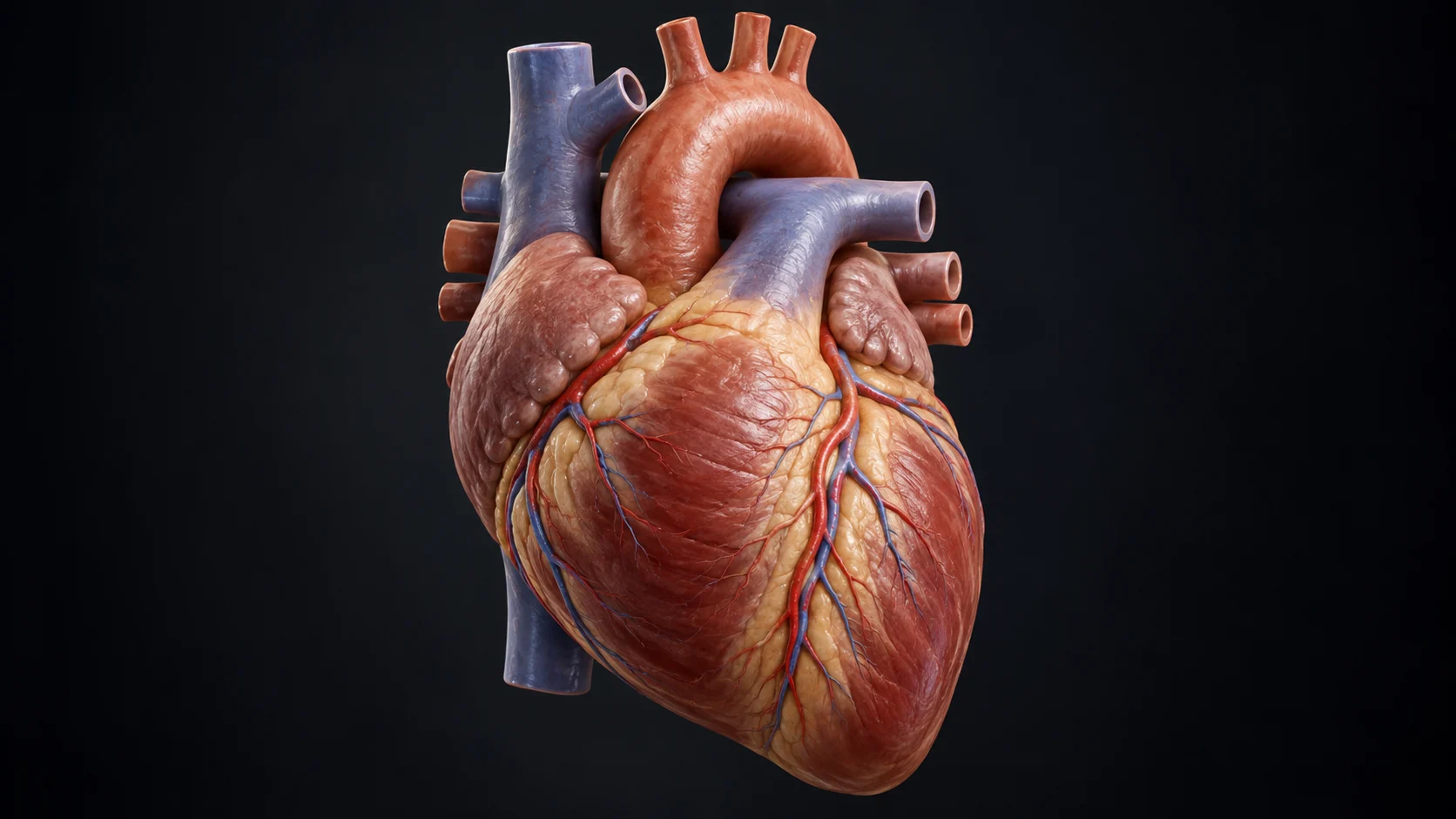
Heart Anatomy 101
Q: What kind of organ is the human heart?
A: A muscular pump.
Q: Where is the heart located in the chest?
A: Behind the sternum, between the lungs, with most of it slightly left of center.
Q: How many chambers does a human heart have?
A: Four.
Q: What are the two upper chambers called?
A: The atria.
Q: What are the two lower chambers called?
A: The ventricles.
Q: What wall separates the right and left sides of the heart?
A: The septum.
Q: What is the pointed lower tip of the heart called?
A: The apex.
Q: What broad upper portion of the heart is called the base?
A: The area where the great vessels attach.
Q: What is the thick muscular middle layer of the heart wall?
A: The myocardium.
Q: What smooth membrane lines the chambers of the heart?
A: The endocardium.
Q: What thin outer layer covers the heart's surface?
A: The epicardium.
Q: What double-walled sac surrounds the heart?
A: The pericardium.
Q: What does fluid within the pericardial sac help reduce?
A: Friction as the heart beats.
Q: Which chamber has the thickest muscular wall?
A: The left ventricle.
Q: Why is the left ventricle more muscular than the right ventricle?
A: It pumps blood through the entire body.
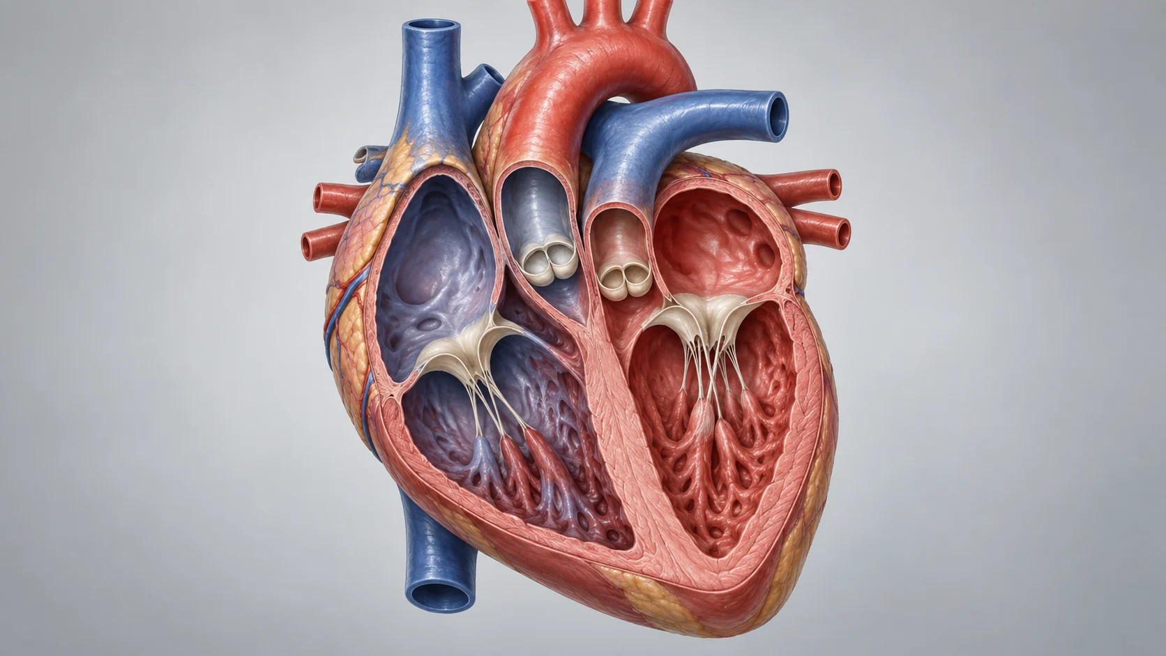
Following the Blood
Q: Which chamber receives oxygen-poor blood returning from the body?
A: The right atrium.
Q: Which chamber pumps oxygen-poor blood toward the lungs?
A: The right ventricle.
Q: Which chamber receives oxygen-rich blood from the lungs?
A: The left atrium.
Q: Which chamber pumps oxygen-rich blood into the aorta?
A: The left ventricle.
Q: What two large veins return blood from the body to the right atrium?
A: The superior and inferior vena cava.
Q: Which vena cava returns blood from the head, neck, and upper body?
A: The superior vena cava.
Q: Which vena cava returns blood from below the diaphragm?
A: The inferior vena cava.
Q: What vessel carries blood from the right ventricle toward the lungs?
A: The pulmonary trunk.
Q: What arteries carry oxygen-poor blood into the lungs?
A: The pulmonary arteries.
Q: What vessels return oxygen-rich blood from the lungs to the heart?
A: The pulmonary veins.
Q: How many pulmonary veins usually enter the left atrium?
A: Four.
Q: What is the body's largest artery?
A: The aorta.
Q: What circuit carries blood between the heart and lungs?
A: Pulmonary circulation.
Q: What circuit carries blood between the heart and the rest of the body?
A: Systemic circulation.
Q: Do all arteries carry oxygen-rich blood?
A: No.
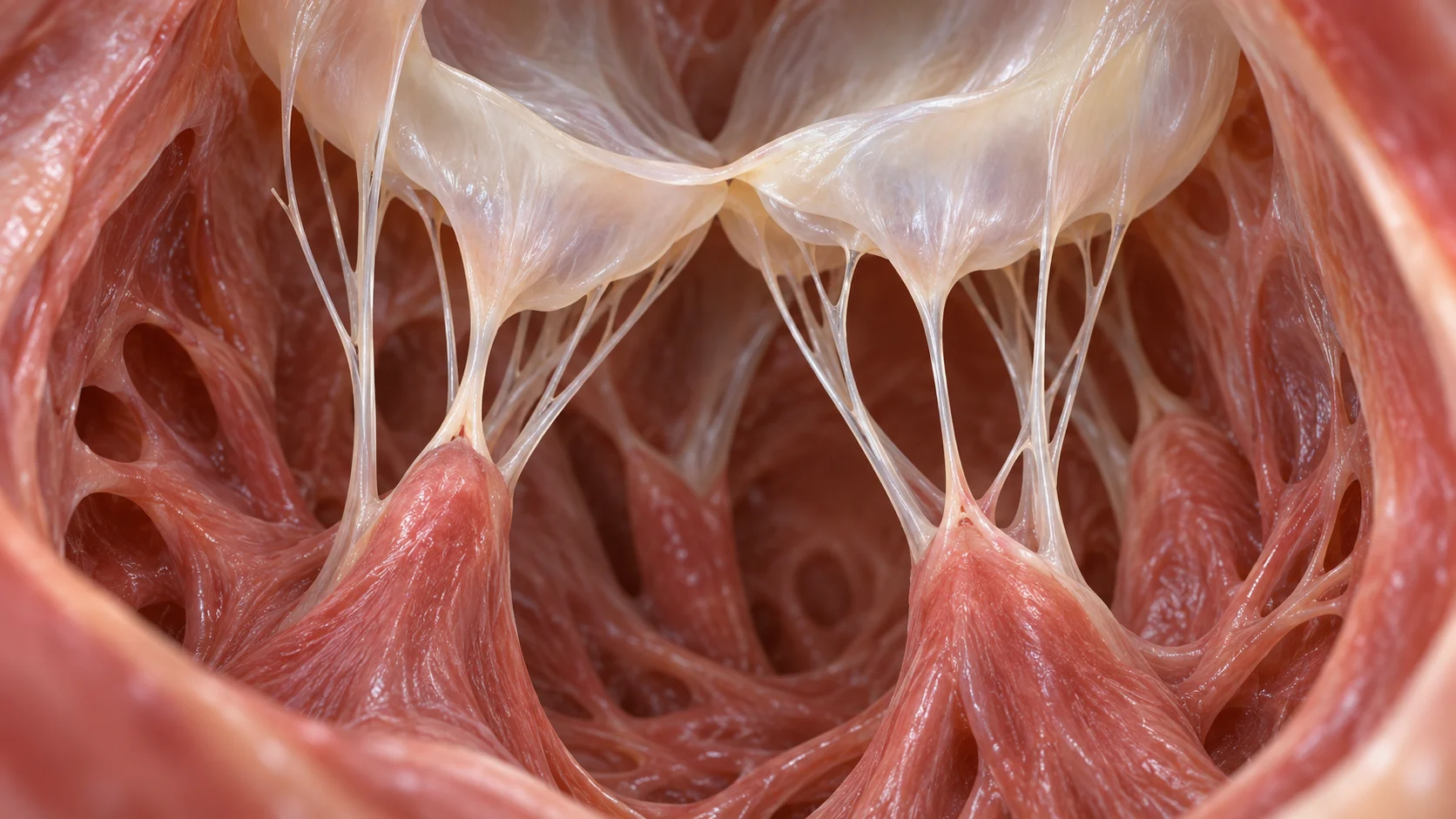
Valves Keep Traffic Moving
Q: How many valves are in the heart?
A: Four.
Q: What valve lies between the right atrium and right ventricle?
A: The tricuspid valve.
Q: What valve lies between the left atrium and left ventricle?
A: The mitral valve.
Q: What other name is used for the mitral valve?
A: The bicuspid valve.
Q: What valve controls blood leaving the right ventricle?
A: The pulmonary valve.
Q: What valve controls blood leaving the left ventricle?
A: The aortic valve.
Q: What collective name is given to the tricuspid and mitral valves?
A: Atrioventricular valves.
Q: What collective name is given to the pulmonary and aortic valves?
A: Semilunar valves.
Q: What is the main job of all four heart valves?
A: To keep blood moving in the proper direction.
Q: What flap-like parts of a valve open and close?
A: Cusps, or leaflets.
Q: How many cusps does the tricuspid valve have?
A: Three.
Q: How many cusps does the mitral valve have?
A: Two.
Q: What tough cords anchor the atrioventricular valve leaflets?
A: Chordae tendineae.
Q: What ventricular muscles tighten the chordae tendineae during contraction?
A: Papillary muscles.
Q: Do chordae tendineae attach to the aortic and pulmonary valves?
A: No.
The Heart's Electrical System
Q: What cluster of cells normally starts each heartbeat?
A: The sinoatrial node.
Q: What familiar nickname is given to the sinoatrial node?
A: The heart's natural pacemaker.
Q: In which chamber is the sinoatrial node located?
A: The right atrium.
Q: What node briefly delays the electrical signal before it reaches the ventricles?
A: The atrioventricular node.
Q: What conduction pathway carries the signal from the atrioventricular node toward the ventricles?
A: The bundle of His.
Q: Into what two pathways does the bundle of His divide?
A: Right and left bundle branches.
Q: What fibers spread the electrical impulse through the ventricular muscle?
A: Purkinje fibers.
Q: What does the heart's electrical signal make cardiac muscle cells do?
A: Contract in a coordinated sequence.
Q: What test records the heart's electrical activity from the body surface?
A: An electrocardiogram.
Q: What abbreviation is commonly used for electrocardiogram?
A: ECG or EKG.
Q: Which ECG wave represents atrial depolarization?
A: The P wave.
Q: Which ECG complex represents ventricular depolarization?
A: The QRS complex.
Q: Which ECG wave represents ventricular repolarization?
A: The T wave.
Q: What term describes the regular pattern of the heartbeat?
A: Heart rhythm.
Q: Can the heart generate its own electrical impulses without direct commands for every beat from the brain?
A: Yes.
A Beat-by-Beat Cycle
Q: What phase of the cardiac cycle involves chamber contraction?
A: Systole.
Q: What phase of the cardiac cycle involves chamber relaxation and filling?
A: Diastole.
Q: What do the two numbers in a blood pressure reading represent?
A: Systolic and diastolic pressure.
Q: What does heart rate count?
A: Heartbeats per minute.
Q: What is the amount of blood pumped by one ventricle in one beat called?
A: Stroke volume.
Q: What is the amount of blood pumped by one ventricle per minute called?
A: Cardiac output.
Q: What two measurements are multiplied to calculate cardiac output?
A: Heart rate and stroke volume.
Q: What fraction describes how much blood a ventricle ejects with each contraction?
A: Ejection fraction.
Q: What is the pulse?
A: The pressure wave created as the heart pumps blood through arteries.
Q: Which artery at the wrist is commonly used to check a pulse?
A: The radial artery.
Q: Which artery in the neck can be used to check a pulse?
A: The carotid artery.
Q: What makes the first heart sound, often written as lub?
A: Closure of the mitral and tricuspid valves.
Q: What makes the second heart sound, often written as dub?
A: Closure of the aortic and pulmonary valves.
Q: What instrument is used to listen to heart sounds?
A: A stethoscope.
Q: During exercise, why does cardiac output normally rise?
A: Working tissues need more oxygen and nutrients.
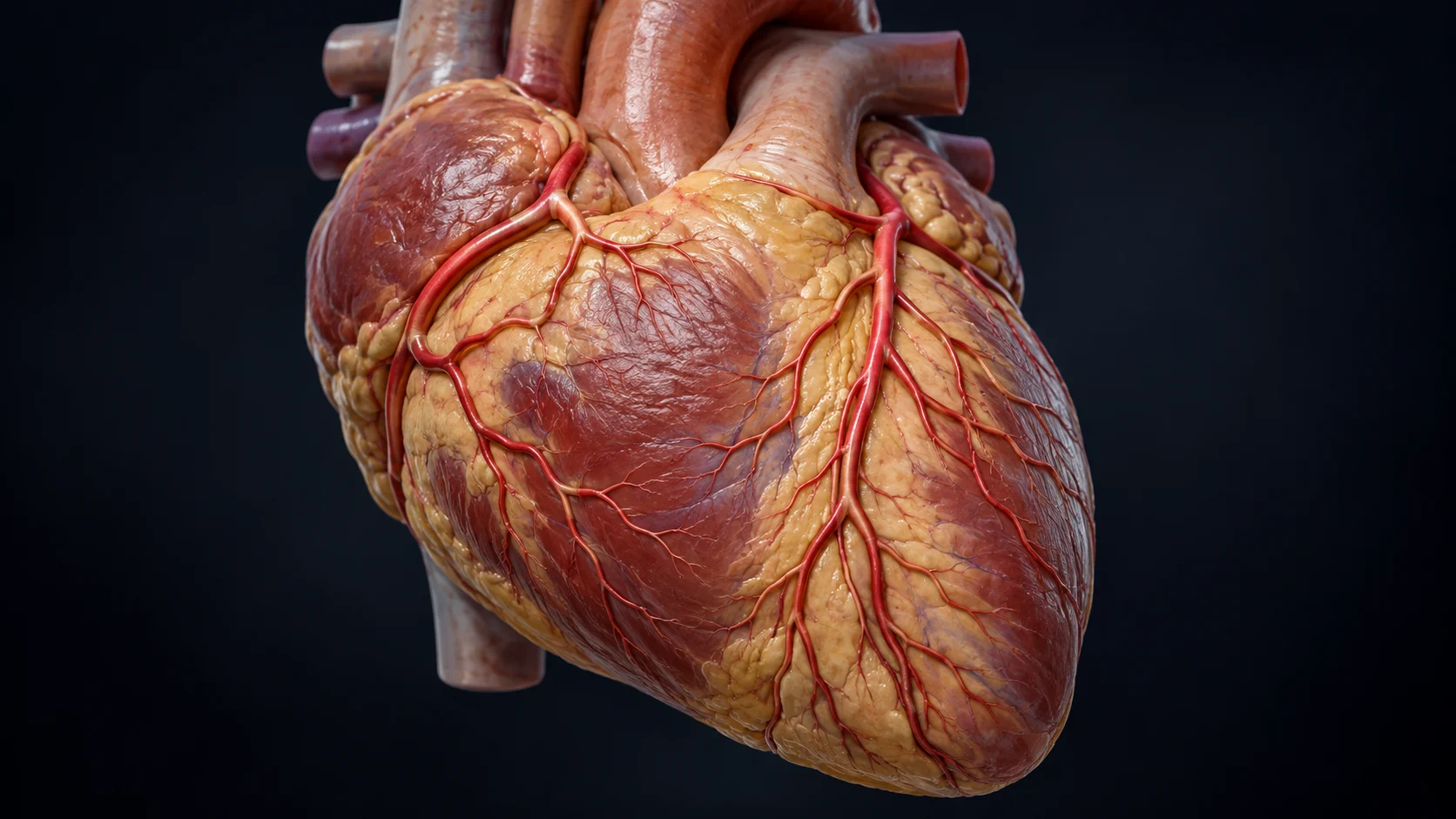
Feeding the Heart Muscle
Q: What vessels supply oxygen-rich blood to the heart muscle itself?
A: The coronary arteries.
Q: From what major artery do the coronary arteries arise?
A: The aorta.
Q: What are the two main coronary arteries?
A: The right and left coronary arteries.
Q: What major branch of the left coronary artery runs down the front of the heart?
A: The left anterior descending artery.
Q: What branch of the left coronary artery curves around the left side of the heart?
A: The circumflex artery.
Q: What network returns oxygen-poor blood from the heart muscle?
A: The cardiac veins.
Q: What large venous channel empties blood from the heart muscle into the right atrium?
A: The coronary sinus.
Q: Why does the heart muscle need its own blood supply?
A: It needs oxygen and nutrients to keep contracting.
Q: What tiny vessels allow exchange between blood and heart tissue?
A: Capillaries.
Q: What protein in red blood cells carries most oxygen?
A: Hemoglobin.
Q: Does blood inside a heart chamber directly nourish all of the thick myocardium?
A: No.
Q: During which phase does much of the left ventricular coronary blood flow occur?
A: Diastole.
Q: What waste gas does coronary blood carry away from heart muscle cells?
A: Carbon dioxide.
Q: What process lets oxygen move from capillary blood into heart tissue?
A: Diffusion.
Q: What molecule made in cells provides immediate usable energy for contraction?
A: ATP.
Before Birth and Across a Lifetime
Q: Is the heart among the first functional organs to develop in an embryo?
A: Yes.
Q: What temporary fetal opening connects the right and left atria?
A: The foramen ovale.
Q: What fetal vessel connects the pulmonary artery to the aorta?
A: The ductus arteriosus.
Q: Why does fetal circulation largely bypass the lungs?
A: Oxygen comes from the placenta rather than from breathing air.
Q: What vessel carries oxygen-rich blood from the placenta toward the fetus?
A: The umbilical vein.
Q: What vessels carry blood from the fetus toward the placenta?
A: The umbilical arteries.
Q: What change at birth helps send more blood through the newborn's lungs?
A: The lungs expand and pulmonary resistance falls.
Q: What normally happens to the foramen ovale after birth?
A: It closes.
Q: What ligament remains after the ductus arteriosus closes?
A: The ligamentum arteriosum.
Q: Is a newborn's resting heart rate generally faster or slower than an adult's?
A: Faster.
Q: What happens to the heart's size as the body grows from infancy to adulthood?
A: It increases.
Q: What nervous system branch usually speeds the heart during stress or activity?
A: The sympathetic nervous system.
Q: What nervous system branch usually slows the resting heart rate?
A: The parasympathetic nervous system.
Q: What major nerve carries parasympathetic signals to the heart?
A: The vagus nerve.
Q: Does the heart stop working while a person sleeps?
A: No.
Seeing, Measuring, and Understanding the Heart
Q: What imaging test uses sound waves to create moving pictures of the heart?
A: An echocardiogram.
Q: What type of echocardiography shows the direction and speed of blood flow?
A: Doppler echocardiography.
Q: What imaging method uses magnets and radio waves to make detailed heart images?
A: Cardiac MRI.
Q: What imaging scan uses X-rays to create cross-sectional views of the heart?
A: Cardiac CT.
Q: What thin tube can be guided through a blood vessel into the heart for testing or treatment?
A: A catheter.
Q: What procedure uses contrast dye and X-rays to view coronary arteries?
A: Coronary angiography.
Q: What wearable device continuously records an ECG for a day or longer?
A: A Holter monitor.
Q: What test measures the heart while a person exercises under supervision?
A: An exercise stress test.
Q: What device delivers timed electrical impulses when the heart's own rhythm is too slow or unreliable?
A: A pacemaker.
Q: What device can deliver a shock to help correct certain dangerous rhythms?
A: A defibrillator.
Q: Who described the circulation of blood driven by the heart in a landmark 1628 work?
A: William Harvey.
Q: What physician developed the stethoscope in the early 1800s?
A: René Laennec.
Q: What does the medical prefix cardio- refer to?
A: The heart.
Q: What does a sphygmomanometer measure?
A: Blood pressure.
Q: Is the familiar heart symbol shaped exactly like the anatomical human heart?
A: No.
You Never Missed a Beat
If you powered through all 120 answers, your trivia rhythm is wonderfully steady. Put the rest of your biology knowledge to work with our science trivia.

Ellie Ewert is the founder and author of RandomTrivia.co, blending her passion for research with years of experience in content creation to deliver accurate, engaging, and well-sourced trivia. Dedicated to providing readers with trustworthy and entertaining facts, she applies meticulous fact-checking and SEO expertise to ensure every article meets the highest standards. Read more about our high standards in our Editorial Guidelines.